
Medical Happenings
Diagnose Early
56 year old lady with recently diagnosed Breast lump 10 days back. Tru cut biopsy suggestive of malignant tumor. On evaluation for secondaries found to have a solitary lesion in her right frontal lobe of brain with mass effect and midline shift of 8mm. Gross total excision of tumor done and patient discharged on second post op day without any deficits. Patient will be started on chemotherapy and radiotherapy in Dharan cancer centre. A holistic management for malignant tumor with high safety and affordability is now available at Dharan hospitals, salem
Periodical Correction & Strapping
8 Months Old Preterm Neonate with Calcaneus Deformity R Foot. A very rare case. Treated by Periodical Correction and Strapping. Today after 3 weeks foot got Corrected with Good Movements. Thanks to Dr.Pramya & Dr.Kuralvanan for giving this Opportunity.
FREE FLAP
Tissue transplantation from one part of body to other, to reconstruct the defect. Tissue is completely detached from the body. After tissue transfer, circulation is reestablished by joining the vein and artery.
Advantages
- Cosmesis (Positive Aesthetic & Functional outcome)
- Large defect can be corrected.
- Can use all type of tissues ( Bone, Soft Tissues, Skin)
- Lower morbidity of Donor Site.
Tracheal Stenosis
An older man was suffering from critical Tracheal Stenosis due to Oesophageal Cancer.
Our Team performed a delicate and complicated procedure called "Mechanical debulking of endotracheal Mass & Placement of Tracheobronchial Stent."
Post Surgery X-rays showed the precise placement of the tracheobronchial stenting. And the gentleman was on his way to a full recovery.
A Rare Case of Spinal AVM
70 Year old female had a difficulty in walking for past 1 year. She came to us in completely bedridden and inability to walk since last 1-2 weeks. She underwent MRI which showed Spinal Myelopathy due to Spinal Vascular Malformation. Our Interventional Radiologist performed Spinal Angiogram which showed Filum Terminale arteriovenous fistula causing Spinal Cord Congestion and Edema. After multi-disciplinary discussion she underwent surgical exclusion of the Filum Terminale Arteriovenous Fistula based on the anatomical localisation in Spinal Angiogram. Post-surgery, she was able to walk and regained her mobility. Filum Terminale Arteriovenous Fistula is rare condition and reported less than 100 cases in literature. Spinal Myelopathy due to Spinal Vascular Malformation can be completely cured if and treated early and appropriately.
A Case of Ruptured intra-cranial Aneurysm
49 year old lady came to emergency in an unconscious state with two days history of severe headache and two episodes of vomiting on the day of admission. Her CT brain revealed a right Sylvian fissure grade III Sub arachnoid hemorrhage. An aneurysm rupture was suspected and patient underwent Digital subtraction angiography. Angiogram revealed a multiple lobed right Middle cerebral artery bifurcation aneurysm.
She underwent clipping of aneurysm and post clipping she was monitored in hospital for vasospasm for ten days. Her post-operative period was uneventful and she was discharged on 11th post op day with no weakness of limbs or speech difficulties. She was able to return to all her routine activities the very next day.
Our teams of critical care Physicians, Interventional Radiologist and Neurologist / Neurosurgeons are able to provide quality care 24/7 at an affordable cost to the people of Salem at par with care available in METRO Cities.
Preterm Neonate with Rigid Right Club Foot
A baby born with a rigid club foot in the right leg, underwent Postero Medial Soft Tissue release operation done by Ortho Surgeon Dr. M.C.R. Nakkeeran. Post Surgery baby was fine, and reviwed periodically. Now walks well as shown in image.
Crush Injury Right Leg Having Fracture Both Bones
20 years young man came under emergency condition Crush injury Right leg having fracture both bones. Wound debridement and external fixator application followed by split skin grafting done by Plastic Surgeon Dr. S. Dhanaraju. External fixator removed and Interlocking nailing Right Tibia. Physiotherapy was given to the patient and reviewed periodically. After wound healing and fractures re-union now the patient walks well.
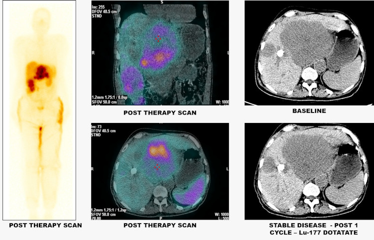
Molecular Theranostics for treatment of Metastic Neuroendocrine Tumour
A 64-year-old gentleman presented with multiple liver lesions. Biopsy revealed a grade II metastatic neuroendocrine tumor Ga-68 DOTANOC PET/CT showed only secondaries in the liver with high somatostatin receptor positivity in all the lesions and no obvious primary. Patient was put on first line of management with octreotide long acting repeatable. But he was having progressive disease with increase in size of the liver lesions.
Further patient was taken up for PRRT (peptide receptor radionuclide therapy) with Lutetium-177 DOTATATE. It is a specific radioactive molecule that delivers radiation only to this variety of tumours expressing this particular receptor with very limited side effects to the rest of the body as compared to conventional chemotherapy or radiotherapy. In this patient, post 1 cycle of therapy, there is no further increase in size of the lesion with no new lesions, thereby achieving stable disease.
This type of management is available only in very few centers throughout the country and Dharan Hospital is the only center in Salem providing this line of treatment.
Youngest Thrombectomy For Stroke in Salem
A 20 years Male, had left upper limb pain for 4 months and ring finger tip blackening since 20 days. He presented to hospital with stroke symptoms in drowsy state with left hemiplegia (Inability to move left side of body). He underwent CT Angiogram and MRI which showed Embolic occlusion to basilar artery with associated left sub clavian artery occlusion due to Thoracic outlet obstruction. He underwent Emergency Mechanical Thrombectomy for basilar artery with successful recovery. This is a rare and unusual case of major stroke. This is the youngest patient treated by Mechanical Thrombectomy in this region.
Spinal Stabilization for Paraplegia in Chronic Liver Disease / Hepatic Failure Patient
44 year old male in liver failure with L1 vertebral body fracture following a trivial fall. He was paraplegia post fall and referred to us for surgical stabilisation of the spine. Patient had a platelet of 30,000 and Hb of 8 due to Chronic Liver Disease and related portal hypertension, splenomegaly. After explaining in detail regarding the complications and post op liver failure he was taken up for surgery. Since it was a transition segment 4 level pedicle screws was planned.
Case was discussed with Anesthesia team and Gastroenterologist. Since there was splenomegaly and patient already in liver dysfunction any platelet transfusion will not improve his levels. So, he was planned for surgery with 2 single donor platelet and 4 fresh frozen plasma on flow. The procedure was completed in 1.30 hrs and patient extubated by 2 hrs and shifted to ward after observing for any liver failure signs. He went in for liver failure on second post op day and platelets dropped to 10k and bilirubin to 15 and Hb to 6. All these were previously anticipated and managed very well by Dr. Gopalakrishnan. He was mobilised and weight bearing exercises started on 5th Post Operative Delirium.. Now the patient is walking on his own. We wanted to highlight the outcome of this patient which was possible only due to the team effort.
Balloon-occluded Retrograde Transvenous Obliteration of Varices
A 60 year female had chronic liver disease with portal hypertension.Recurrent Upper Gastrointestinal Series bleed due to bleeding from large gastric varices. UGI scopy confirmed the bleeding from gastric varices. She underwent BRTO with successful control of bleeding.
We enter from Right femoral vein - IVC - renal vein -Gastro renal shunt. Occluded gastro renal shunt using balloon/coil/plug and obliterate the gastric varices with sclerosant mixture. We study the feasibility,afferents ,efferents of varices using Contrast-enhanced computed tomography which guides for treatment. Gastric varices are predominantly submucosal unlike esophageal varices .So watever seen in endoscopy is only tip of iceberg and hence the complete elimination of gastric varices is difficult by endoscopic methods and more chances of recurrence. Thanks to the Gastroenterologist team.
Diagnose Early
56 year old lady with recently diagnosed Breast lump 10 days back. Tru cut biopsy suggestive of malignant tumor. On evaluation for secondaries found to have a solitary lesion in her right frontal lobe of brain with mass effect and midline shift of 8mm. Gross total excision of tumor done and patient discharged on second post op day without any deficits. Patient will be started on chemotherapy and radiotherapy in Dharan cancer centre. A holistic management for malignant tumor with high safety and affordability is now available at Dharan hospitals, salem

Periodical Correction & Strapping
8 Months Old Preterm Neonate with Calcaneus Deformity R Foot. A very rare case. Treated by Periodical Correction and Strapping. Today after 3 weeks foot got Corrected with Good Movements. Thanks to Dr.Pramya & Dr.Kuralvanan for giving this Opportunity.

FREE FLAP
Tissue transplantation from one part of body to other, to reconstruct the defect. Tissue is completely detached from the body. After tissue transfer, circulation is reestablished by joining the vein and artery.
Advantages
- Cosmesis (Positive Aesthetic & Functional outcome)
- Large defect can be corrected.
- Can use all type of tissues ( Bone, Soft Tissues, Skin)
- Lower morbidity of Donor Site.

Tracheal Stenosis
An older man was suffering from critical Tracheal Stenosis due to Oesophageal Cancer.
Our Team performed a delicate and complicated procedure called "Mechanical debulking of endotracheal Mass & Placement of Tracheobronchial Stent."
Post Surgery X-rays showed the precise placement of the tracheobronchial stenting. And the gentleman was on his way to a full recovery.

A Rare Case of Spinal AVM
70 Year old female had a difficulty in walking for past 1 year. She came to us in completely bedridden and inability to walk since last 1-2 weeks. She underwent MRI which showed Spinal Myelopathy due to Spinal Vascular Malformation. Our Interventional Radiologist performed Spinal Angiogram which showed Filum Terminale arteriovenous fistula causing Spinal Cord Congestion and Edema. After multi-disciplinary discussion she underwent surgical exclusion of the Filum Terminale Arteriovenous Fistula based on the anatomical localisation in Spinal Angiogram. Post-surgery, she was able to walk and regained her mobility. Filum Terminale Arteriovenous Fistula is rare condition and reported less than 100 cases in literature. Spinal Myelopathy due to Spinal Vascular Malformation can be completely cured if and treated early and appropriately.

A Case of Ruptured intra-cranial Aneurysm
49 year old lady came to emergency in an unconscious state with two days history of severe headache and two episodes of vomiting on the day of admission. Her CT brain revealed a right Sylvian fissure grade III Sub arachnoid hemorrhage. An aneurysm rupture was suspected and patient underwent Digital subtraction angiography. Angiogram revealed a multiple lobed right Middle cerebral artery bifurcation aneurysm.
She underwent clipping of aneurysm and post clipping she was monitored in hospital for vasospasm for ten days. Her post-operative period was uneventful and she was discharged on 11th post op day with no weakness of limbs or speech difficulties. She was able to return to all her routine activities the very next day.
Our teams of critical care Physicians, Interventional Radiologist and Neurologist / Neurosurgeons are able to provide quality care 24/7 at an affordable cost to the people of Salem at par with care available in METRO Cities.

Preterm Neonate with Rigid Right Club Foot
A baby born with a rigid club foot in the right leg, underwent Postero Medial Soft Tissue release operation done by Ortho Surgeon Dr. M.C.R. Nakkeeran. Post Surgery baby was fine, and reviwed periodically. Now walks well as shown in image.

Crush Injury Right Leg Having Fracture Both Bones
20 years young man came under emergency condition Crush injury Right leg having fracture both bones. Wound debridement and external fixator application followed by split skin grafting done by Plastic Surgeon Dr. S. Dhanaraju. External fixator removed and Interlocking nailing Right Tibia. Physiotherapy was given to the patient and reviewed periodically. After wound healing and fractures re-union now the patient walks well.

Molecular Theranostics for treatment of Metastic Neuroendocrine Tumour
A 64-year-old gentleman presented with multiple liver lesions. Biopsy revealed a grade II metastatic neuroendocrine tumor Ga-68 DOTANOC PET/CT showed only secondaries in the liver with high somatostatin receptor positivity in all the lesions and no obvious primary. Patient was put on first line of management with octreotide long acting repeatable. But he was having progressive disease with increase in size of the liver lesions. Further patient was taken up for PRRT (peptide receptor radionuclide therapy) with Lutetium-177 DOTATATE. It is a specific radioactive molecule that delivers radiation only to this variety of tumours expressing this particular receptor with very limited side effects to the rest of the body as compared to conventional chemotherapy or radiotherapy. In this patient, post 1 cycle of therapy, there is no further increase in size of the lesion with no new lesions, thereby achieving stable disease. This type of management is available only in very few centers throughout the country and Dharan Hospital is the only center in Salem providing this line of treatment.
Youngest Thrombectomy For Stroke in Salem
A 20 years Male, had left upper limb pain for 4 months and ring finger tip blackening since 20 days. He presented to hospital with stroke symptoms in drowsy state with left hemiplegia (Inability to move left side of body). He underwent CT Angiogram and MRI which showed Embolic occlusion to basilar artery with associated left sub clavian artery occlusion due to Thoracic outlet obstruction. He underwent Emergency Mechanical Thrombectomy for basilar artery with successful recovery. This is a rare and unusual case of major stroke. This is the youngest patient treated by Mechanical Thrombectomy in this region.

Spinal Stabilization for Paraplegia in Chronic Liver Disease / Hepatic Failure Patient
44 year old male in liver failure with L1 vertebral body fracture following a trivial fall. He was paraplegia post fall and referred to us for surgical stabilisation of the spine. Patient had a platelet of 30,000 and Hb of 8 due to Chronic Liver Disease and related portal hypertension, splenomegaly. After explaining in detail regarding the complications and post op liver failure he was taken up for surgery. Since it was a transition segment 4 level pedicle screws was planned.
Case was discussed with Anesthesia team and Gastroenterologist. Since there was splenomegaly and patient already in liver dysfunction any platelet transfusion will not improve his levels. So, he was planned for surgery with 2 single donor platelet and 4 fresh frozen plasma on flow. The procedure was completed in 1.30 hrs and patient extubated by 2 hrs and shifted to ward after observing for any liver failure signs. He went in for liver failure on second post op day and platelets dropped to 10k and bilirubin to 15 and Hb to 6. All these were previously anticipated and managed very well by Dr. Gopalakrishnan. He was mobilised and weight bearing exercises started on 5th Post Operative Delirium.. Now the patient is walking on his own. We wanted to highlight the outcome of this patient which was possible only due to the team effort.

Balloon-occluded Retrograde Transvenous Obliteration of Varices
A 60 year female had chronic liver disease with portal hypertension.Recurrent Upper Gastrointestinal Series bleed due to bleeding from large gastric varices. UGI scopy confirmed the bleeding from gastric varices. She underwent BRTO with successful control of bleeding.
We enter from Right femoral vein - IVC - renal vein -Gastro renal shunt. Occluded gastro renal shunt using balloon/coil/plug and obliterate the gastric varices with sclerosant mixture. We study the feasibility,afferents ,efferents of varices using Contrast-enhanced computed tomography which guides for treatment. Gastric varices are predominantly submucosal unlike esophageal varices .So watever seen in endoscopy is only tip of iceberg and hence the complete elimination of gastric varices is difficult by endoscopic methods and more chances of recurrence. Thanks to the Gastroenterologist team.